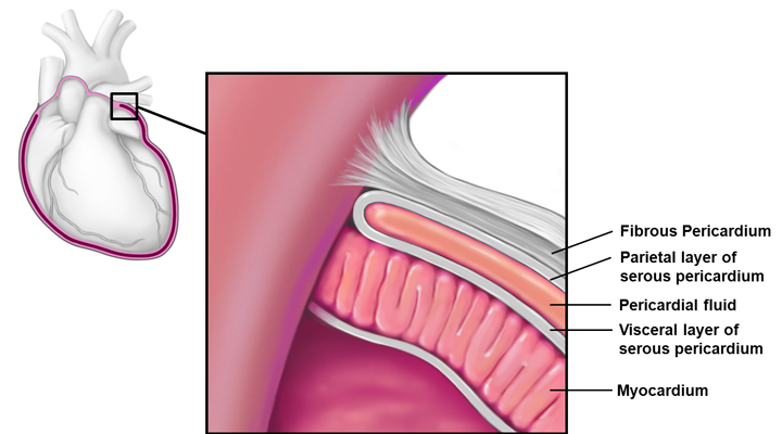
The layers have a small amount of fluid between them to prevent friction when the heart. Sympathetic Fibers increase the rate and force of contraction travel to the heart from the cervical and upper thoracic sympathetic chain of ganglia and innervate the visceral layer of serous pericardium just like the sympathetic.
Pin On Pdf Download
The pericardium is a thin sac that surrounds your heart.
Heart pericardium. Visceral and parietal are separated by the pericardial cavity which contains 20 to 60 mL of the plasma ultrafiltrate. The heart and pericardium are situated behind the sternum breastbone in a position in the middle of the chest cavity known as the mediastinum. Pericardium is a thin conical fibroelastic sac or a membrane surrounding hearts and its great vessels.
It protects and lubricates your heart and keeps it in place within your chest. Heart and Pericardium Video Notes. Pericarditis is inflammation of the pericardium a sac-like structure with two thin layers of tissue that surround the heart to hold it in place and help it work.
The pericardium acts as mechanical protection for the heart and big vessels and a lubrication to reduce friction between the heart. The heart and pericardium This article describes the normal anatomy of the heart and pericardium. The pericardium protects the heart from emotional trauma constricts the chest to protect the heart and helps to express the joy of the heart.
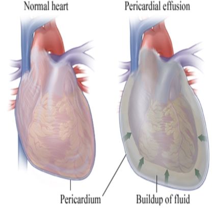
As cardiac and thoracic surgery continue to get more specialized and t. A pericardial effusion is excess fluid between the heart and the sac surrounding the heart known as the pericardium. Heart rate arise as branches of the vagus nerve in the neck and thorax and innervate the visceral layer of serous pericardium.
A small amount of fluid keeps the layers separate so theres less friction between them as the heart beats. Pericarditis is the inflammation of the pericardium a thin two-layered sac that surrounds your heart. The heart and its pericardium make up the contents of the middle mediastinum The left and right phrenic nerves and the pericardiacophrenic vessels lie to the left and right of the pericardium respectively.
Included is a detailed description of the pericardium mediastinal nerves cardiac chambers valves coronary arteries and veins and the conduction tissues. In scientific terms the pericardium is a fibro-serous fluid-filled sack that surrounds the muscular body of the heart and the roots of the great vessels the aorta pulmonary artery pulmonary veins and the superior and inferior vena cavae. Back to the mediastinum and the heart we take a look at an imaginary pericardium.
The fibrous pericardium consists of thick fibrous connective tissue and it defines the borders of the middle mediastinum. Problems can occur when the pericardium. The pericardium is a fibro-serous sac that encloses the heart.
The pericardium is the membrane that encloses the heart and the roots of the major heart vessels consisting of an outer fibrous layer fibrous pericardium and an inner double serous membrane layer serous pericardium. Most are not harmful but they. What are its layers where is it and why is it thereDaily Anatomy AppFo.
The pericardium is the fluid-filled sac that surrounds the heart and the proximal ends of the aorta venae cavae and the pulmonary artery. The two layers of serous pericardium. The pericardium protects the heart from potential damage caused from the strong fluctuations in energy caused by emotional ups and downs of the day.
Various functions of pericardium include preventing dislocation of heart by maintaining its original position providing mechanical support to the heart and its great vessels and act as a lubrication to lessen friction between the heart and. The pericardium is a thin two-layered fluid-filled sac that covers the outer surface of the heart. It provides lubrication for the heart shields the heart from infection and malignancy and contains the heart.

19 6 Pericardium The Protective Layers Of The Heart Include The Pericardial Sac Composed Of An Outer Anatomy Models Human Anatomy And Physiology Heart Anatomy

Pericardium Serous Membranes Visceral Parietal Layers Medical Coding Human Anatomy And Physiology Serous Membrane

Structure Of The Heart Heart Structure Arteries And Veins Anatomy And Physiology

Layers Of The Pericardium Heart Wall And Spiral Arrangement Cardiovascular System Biology Lessons Medical Anatomy

Layers Of The Heart And Pericardium Nursing Videos Anatomy And Physiology Cardiovascular System

Layers Of The Heart Muscle And Pericardium The Section Of The Heart Wall Shows The Fibrous Pericardium The Par Heart Disorders Medical Knowledge Heart Muscle

Pericardium Epicardium Myocardium Endocardium Google Search Mitral Valve Mitral Valve Prolapse Enlarged Heart

Pericarditis Is Inflammation Of The Pericardium The Sac Which Surrounds The Heart Pericarditis Causes Chest Pains Per Best Hospitals Hospital Health Facts
5 Home Remedies For Pericarditis Pericardial Effusion Cardiac Nurse

Pin Em Top Health Topics Blood Heart And Circulation

Pericardium And Heart Pericardium The Heart Sac Anatomy And Physiology Heart Flashcards

Pin On Anatomy

Pericarditis Inflammation Of The Heart Tissue Http Www Nlm Nih Gov Medlineplus Ency Article 0 Cardiac Nursing Inflammation Of The Heart Heart Transplant

Pericardium

Pin On Pdf Download

The Pericardium The Pericardium Is A Double Walled Sac That Contains The Heart The Pericardial Sac Is Cardiovascular Nursing Cardiac Nursing Med School Study

A Heart With Pericarditis And A Normal Heart Heart Conditions Pericardial Effusion Heart Attack Symptoms

The Pericardium Is A Double Walled Sac That Encloses The Heart Between The Visceral And Parietal Membranes I Pericardial Effusion Medical Photos Heart Anatomy

Pericarditis Pericardial Effusion Amp Cardiac Tamponade Bmh Tele Pericardial Effusion Cardiac Cardiac Nursing

